Sir Wilfrid Le Gros Clark's neuroanatomical teaching collection
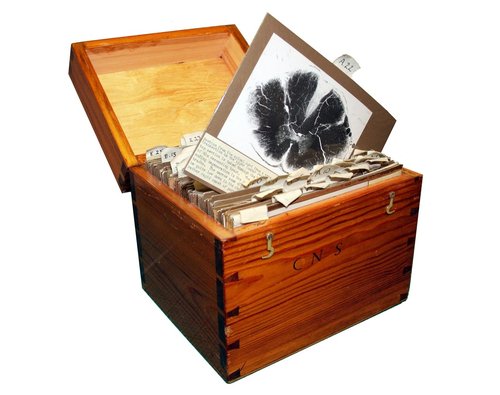
A collection of historic teaching slides preserved by the Department of Physiology, Anatomy and Genetics (DPAG) at the University of Oxford offers fresh insight into the development of modern neuroscience and the enduring educational legacy of pioneering anatomist Sir Wilfrid Edward Le Gros Clark.
In a new paper published in the Journal of Anatomy, a group of undergraduate and graduate students from St John’s College examined selected specimens from Le Gros Clark’s neuroanatomical collection, which remains in the care of DPAG. https://doi.org/10.1111/joa.70235.
Le Gros Clark (1895–1971) was an anatomist, physician and evolutionary biologist whose work helped shape twentieth-century neuroanatomy, primate biology and medical education. He was also instrumental in exposing the Piltdown Man remains as a scientific fraud.
The study explores teaching slides illustrating conditions including tabes dorsalis, multiple sclerosis – historically termed disseminated sclerosis – amyotrophic lateral sclerosis and syringomyelia, as well as the nervus conarii, a structure associated with the pineal gland.
Le Gros Clark's approach to teaching was as deliberate as his approach to research. In the preface to The Tissues of the Body (1939), the introductory textbook that grew out of his Oxford lectures, he argued that such a work should be not merely informative but also 'intellectually stimulating', and that students should be brought from the earliest stage of their training to vantage points from which the advancing front of anatomical research could be seen. Anatomy, on this view, was not a settled body of fact to be committed to memory, but an active field of inquiry into which students were to be inducted directly.
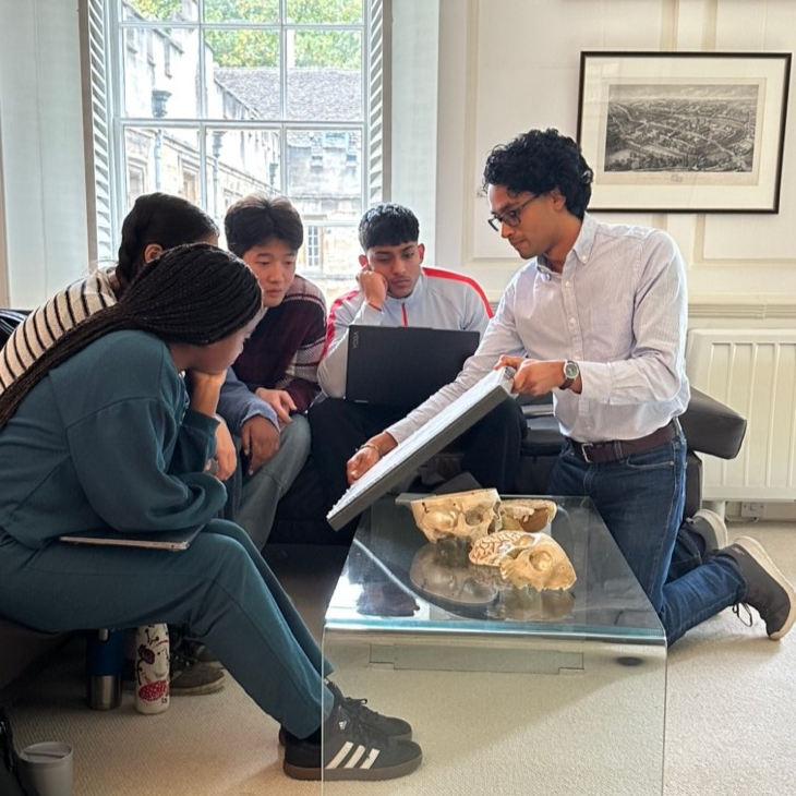 Kush Kale demonstrating for the second-year medical students at St John’s College
Practical classes involved setting up half a dozen slides from the Le Gros Clark collection under microscopes in designated bays, accompanied by corresponding descriptive cards. These cards often featured photographs or annotated diagrams to help students correlate microscopic features with functional anatomy. The application of the anatomical knowledge was reinforced by carefully selected clinical cases. The scanned sections of these cases are still used today in neuroanatomy teaching via computer‐assisted teaching material at the University of Oxford.
By re-examining these specimens in the light of present-day neuroscience, the authors show how Le Gros Clark’s interpretations anticipated concepts that remain central to understanding the nervous system. The collection also reveals his distinctive approach to teaching: encouraging students to observe closely, interpret what they saw and relate anatomical structures to their functions in health and disease.
Professor Zoltán Molnár of St John’s College and Department of Physiology, Anatomy and Genetics, who supervised the study, said:
'The Le Gros Clark teaching slide collection has scientific and educational value, and it is important that we preserve this historic teaching material. I have been using some of these clinical cases for my tutorial teaching at St John’s and in the computer-assisted learning programs for neuroanatomy practicals for the new generations of medical students. It was a pleasure to work with this highly enthusiastic and brilliant group of undergraduate and graduate students from St John’s on this project. Thanks go to Damion Young and Jon Mason from the Medical Sciences Division Learning Technologies Team for all their help with the online digital display and to Peter Belk from DPAG for archiving.'
The paper brings together seven authors: Shiva A. Nischal, Kush M. Kale, Katherine Tourigny, Mohammed Abuelem, Gretchen Greene, Rachel Walsh and Zoltán Molnár. All seven are affiliated with the College, underlining St John's shared connection to this exploration of Oxford’s scientific and educational heritage. Six of the authors are also affiliated with DPAG, while Katherine Tourigny is affiliated with Oxford’s Department of Clinical Neurosciences.
The paper, “Neuroanatomy education through the years: Revisiting the teaching slides used by Sir Wilfrid Edward Le Gros Clark,” was published online in the Journal of Anatomy on 8 September 2026.
Full paper: Shiva A. Nischal, Kush M. Kale, Katherine Tourigny, Mohammed Abuelem, Gretchen Greene, Rachel Walsh and Zoltán Molnár, 'Neuroanatomy education through the years: Revisiting the teaching slides used by Sir Wilfrid Edward Le Gros Clark', Journal of Anatomy. https://doi.org/10.1111/joa.70235
The digitised collection of Le Gros Clark's teaching material is also available.