The Art of Anatomy
- Date 14 July 2018 - 2.00 p.m. - 25 July 2018 - 7.00 p.m.
- Location The Barn Gallery, St John's College, St Giles
The exhibition explores the continuing relationship between artistic and scientific study, and between artistic practice and our understanding of anatomy. It includes work by current and recent Ruskin students, alongside artistic collaborations between anatomists, scientists and artists.
It will reveal, to both a large audience of international scientists and the wider public, how the scientific study of anatomy can be integrated into a fine arts education, and how scientists now work with artists to create an intelligible visual language.
The exhibition runs from 14 to 25 July, to coincide with the Anatomical Society’s Summer Meeting on 23–25 July 2018, also being held in College.
Opening hours:
Sat
14 July: 2 – 5pm
Sun
15 July: 2 – 5pm
Wed
18 July: 5.30 – 7pm
Thu
19 July: 5.30 – 7pm
Saturday
21 July: 2 – 5pm
Opening Hours during Anatomical Society's Meeting:
Tue 24 July: 6 – 7pm
Wed
25 July: 2 - 7pm
'In medicine anatomy is applied, in art it is transformed', Cecil Erskine
There has been a historic tradition of anatomical study in art academies, but few fine art programmes now offer it as a subject. One of the unique aspects of the undergraduate degree offered at the Ruskin School of Art (Oxford University’s Department of Fine Art) is the first-year course in Anatomy. For more than 40 years, the Department of Physiology, Anatomy and Genetics has generously allowed Ruskin students access to the Dissecting Room.
 It
may be considered anachronistic in a programme of contemporary art practice to
study anatomy (and we believe that the Ruskin is the last art school in Europe
where fine artists - as opposed to medical artists - study directly from
cadavers). But the students respond to the subject beyond the technical and
scientific, producing dynamic work across many media, including sculpture,
textiles, photography, performance and video, as well as drawing.
It
may be considered anachronistic in a programme of contemporary art practice to
study anatomy (and we believe that the Ruskin is the last art school in Europe
where fine artists - as opposed to medical artists - study directly from
cadavers). But the students respond to the subject beyond the technical and
scientific, producing dynamic work across many media, including sculpture,
textiles, photography, performance and video, as well as drawing.
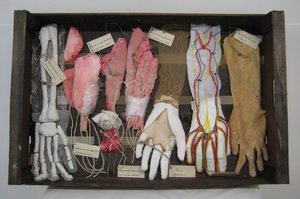
caption: Emily Stevenhagen, Dissected Forearm and Hand, 2018, mixed media: mud, rock, latex, fabric, lentils, thread, wire, skin colour tights and plastic glove. Courtesy the artist.
Dr Sarah Simblet is the Ruskin’s Tutor of Anatomy, and author of Anatomy for the Artist where she writes:
'The study of human anatomy is so much more than the naming of parts and the understanding of their function; it is a celebration of our wondrous physicality in the world. The biological complexity of the body can be weighed against its aesthetic beauty, so that the life that drives us is seen to ripple and pulse above and below the boundary of skin. Art is the perfect tool for revealing such knowledge. Throughout history, artists have used their trained eyes to look deep into our lives and into our very existence. They have given much to the history of medicine and they have also removed the taboo from seeing too much.
... Artists and anatomists have for centuries shared their views and have contributed equally to our knowledge. The excitement and sense of achievement evident in learning about the body is always a palpable delight [...], to catch the perfection of form and reveal its magnificent machinery is highly rewarding. Anatomy is a very potent subject and the subject is us.'
The exhibition includes anatomy works by 15 current and recent Ruskin students, who in their first year are set a two-part assignment by Sarah Simblet:
- to make a drawing
that is larger than themselves, describing the skeleton of an invented
creature; and
- to make an object
that either reflects upon the experience of living inside a body, or choose a
system or part of the body and make an object to explain how it works.
The brief is very fluid, and the students can interpret drawing broadly and imaginatively, without restriction, using any medium from pencil to video; they can take a mechanical approach to the subject, or a philosophical one - or both; they can be humorous or take on a serious issue - or both. Throughout the anatomy course the prime subject is imagination.
 The
student work will be shown alongside artistic collaborations between
anatomists, scientists and artists. Clive Lee is Professor of Anatomy at the
Royal College of Surgeons (RCSI) in Ireland, and at the Royal Hibernian Academy
(RHA). Clive has collaborated with the President of the RHA, Mick O’Dea, and
photographer Amelia Stein RHA, to create a series Portraits of the Anatomist
as a Middle-Aged Man demonstrating
the transformation of anatomy through the artistic process.
The
student work will be shown alongside artistic collaborations between
anatomists, scientists and artists. Clive Lee is Professor of Anatomy at the
Royal College of Surgeons (RCSI) in Ireland, and at the Royal Hibernian Academy
(RHA). Clive has collaborated with the President of the RHA, Mick O’Dea, and
photographer Amelia Stein RHA, to create a series Portraits of the Anatomist
as a Middle-Aged Man demonstrating
the transformation of anatomy through the artistic process.
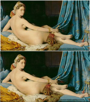
Science and Engineering Academy Award winner, Professor Anil Kokaram, Head of Electronic and Electrical Engineering at Trinity College Dublin (TCD), has made some iconic images anatomically correct. Old Masters Remastered questions the balance between the artistic and the accurate.
caption: Anil Kokaram: above: Jean Auguste Dominiques Ingres, Grande Odalisque, 1814, oil on canvas, Musée du Louvre, Paris (Digital Print / Bridgeman Images); below: Anil Kokaram, Grande Odalisque Remastered, (Digital Print / Anil Kokaram).
Clive, Anil, Mick and colleagues combined to develop a free, online surface anatomy guide - its trailer Anatomists, Engineers & Artists can be seen here – http://www.rcsi.ie/surfaceanatomy
Dr Ciaran Simms, from Mechanical Engineering in TCD and Anatomy in RCSI, has worked with artist Mark Wickham to produce a short film which reflects on the real, the ideal and the safe in motor car accidents – The Biomechanics of Beauty.
In Japan, artists cannot have access to dissection, but Kyoto-based artist Yoshihiro Takada wanted to be able to draw and paint cadavers. So he contacted Professor Mitsuhiro Kawata of the Department of Biological Structure Science at Kyoto University and studied in his Anatomy Laboratory for three years, and has also worked in the Dissecting Room at DPAG under its former Head, Professor John Morris. He has produced new work for this exhibition, combining his explorations of anatomy into his practice in traditional Japanese painting.
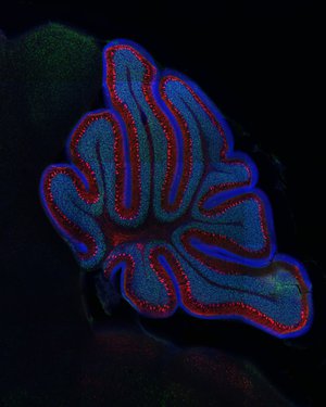 Professor
Zoltán Molnár is Professor of Developmental Neurobiology in
DPAG and Tutorial Fellow of Human Anatomy at St
John’s College. In his laboratory he and his team research the interactions
between the environment and the unfolding genetic programme of brain development,
with special attention to the cerebral cortex. In their studies, they produce
exquisite ‘abstract’ photographs of the brain’s structures, down to the levels
of neurons – taking anatomical imagery down to the cellular.
Professor
Zoltán Molnár is Professor of Developmental Neurobiology in
DPAG and Tutorial Fellow of Human Anatomy at St
John’s College. In his laboratory he and his team research the interactions
between the environment and the unfolding genetic programme of brain development,
with special attention to the cerebral cortex. In their studies, they produce
exquisite ‘abstract’ photographs of the brain’s structures, down to the levels
of neurons – taking anatomical imagery down to the cellular.
caption: Laboratory of Zoltán Molnár, Cerebellum
The idea of this exhibition originated from Professor Molnár, who teamed up with the Ruskin’s Sarah Simblet. St John’s College Research Centre provided support to offer a sabbatical for Professor Clive Lee at St John’s College, to coordinate the exhibition to coincide with the Anatomical Society Summer Meeting he is co-organising with Professor Gavin Clowry (University of Newcastle).
Professor Zoltán Molnár said: 'I am delighted to see the continued interactions between the Ruskin School of Art, the Department of Physiology, Anatomy and Genetics and St John’s College. We are very grateful to St John’s College Research Centre, Cortex Club and British Science Association for their support.'